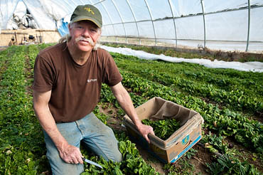
Like a lot of baby boomers, Wisconsin organic farmer Matt Smith grew up watching Lassie save Timmy on television.
Years later, he would have an emergency operation at UW Hospital to save his eyesight. And, yes, there probably was a collie to thank.

Collies were the first model for developing the surgical techniques to fix detached retinas, says Dan Albert, director of the UW–Madison Eye Research Institute.
Albert, a historian of ophthalmology, says that the breeding that created the collie’s long snout also elongated their eyes, leaving them prone to a defect called “collie eye” which makes them myopic and susceptible to detached retinas.

“In the 1950s and 1960s, Dr. Charles Schepens, together with his associates, used collies to develop and refine the techniques of retinal detachment surgery,” says Albert of his former Harvard University colleague, a pioneer in eye surgery.
The retina is the light-sensitive membrane that covers the back wall of the eyeball. It contains the rods and cones that relays visual information through the optic nerve to the brain. In a detached retina, some of the membrane separates from the tissues on the back wall of the eye.
Michael Ip, a UW–Madison eye surgeon, associate professor of ophthalmology and visual sciences and member of the Eye Research Institute, describes it as similar to “a tear in the wallpaper that begins peeling away from the wall.”
Ip says that before Schepens did his pioneering eye research on collies, there was no reliable fix for a detached retina.
“Back then, people would just go blind,” he says. Today, the surgery has a 90 percent likelihood of success; Ip himself does 50-70 such surgeries a year.
But a retinal detachment is still a medical emergency, because the likelihood of restoring vision drops the longer the retina is detached.
Matt Smith’s emergency began on a May morning in 2004, when he woke up and found he had lost much of the sight in his left eye.
About 28,000 people experience a retinal detachment each year in the United States. They can be caused by trauma, such as a blow to the head, or, as in Smith’s case, from years of straining to focus his eyes due to extreme myopia.
“It was really funky, like looking through one of those beaded curtains,” he says. And it couldn’t have come at a worse time: May is the planting season for crops at Smith’s Blue Valley Gardens, located near Blue Mounds west of Madison.
But his local eye doctor took one look into his eye and got on the phone to Ip at UW–Madison. A day later, Smith was under anesthesia at UW Hospital, and Ip was preparing to operate.
Schepens invented many important tools for ophthalmology, including the binocular ophthalmoscope that Smith’s doctor used to look into his eye and diagnose the retinal tear. Schepens also invented and improved a surgical procedure, the “scleral buckle,” in his work on collies during the 1960s.
Ip used that technique to repair Smith’s retinal tear. He sewed a thin silicone band around the circumference of the sclera, or white, of the eye. A buckle on the band pulled tight to create a dimple on the eye wall, bringing the torn retina back in touch with the back wall of the eye. A bubble of gas held the torn piece in place while it healed.
Smith’s eyesight was saved — and after taking it easy for a few weeks while his eye healed, he was back at work on his farm. There was a bonus for the doctor, as well. He and his family are regular patrons of Smith’s produce stand at the Dane County Farmers Market.
“I love his asparagus,” Ip says. “It’s the best asparagus around.”